
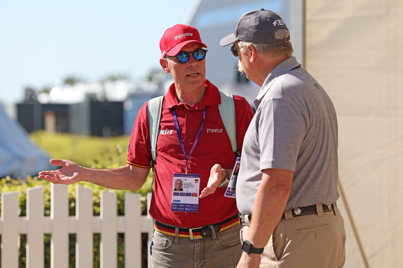
 Jan-Hein Swagemakers is a FEI permitted treating veterinarian, an equine chiropractor and the German team veterinarian since 2009, and runs Tierklinik Lüsche in Germany. Photo © Jenny A Photo for World of Showjumping.
Text © World of Showjumping
“What used to be the standard treatment for these types of diagnoses, namely cervical osteoarthritis or cervical nerve irritation, was injecting the joints. However, only some of the horses improved, while others did not, and some of those who improved saw the issue reoccur,” Dr. Jan-Hein Swagemakers tells World of Showjumping. His new discovery, a minimally invasive endoscopic surgery in the cervical spine, aims to help horses with impinged cervical nerves.
“Now that we have a safe way to do surgery, we can get rid of the underlying problem and not only treat the symptoms,” Swagemakers – a FEI permitted treating veterinarian, an equine chiropractor and the German team veterinarian since 2009, who runs Tierklinik Lüsche in Germany – explains.
A variety of symptoms
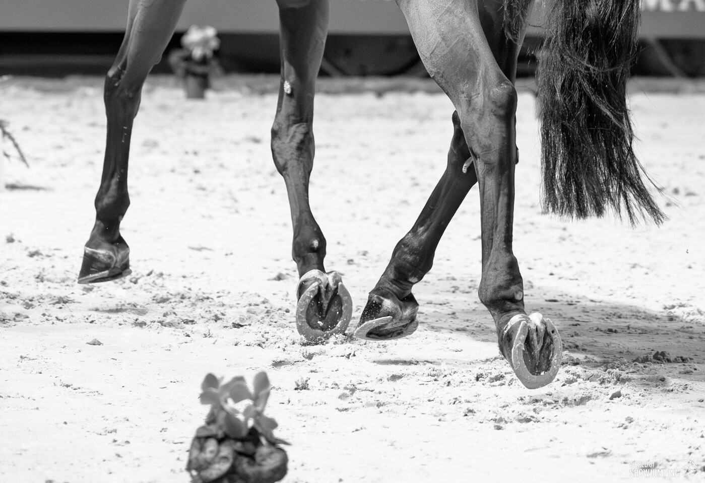 “Some horses can get extremely lame similar to the degree seen in fractures or hoof abscesses – and after 10 minutes, the same horse might start walking normally again," Swagemakers explains. Photo © Jenny A Photo for World of Showjumping.
“If you look at the cross section of the neck, the spinal cord is in the center of the vertebral canal,” Swagemakers explains about the equine cervical spine, which consists of seven vertebrae. “The cervical nerves which are responsible for neck and forelimb movement and sensation have their roots at different levels of the spinal cord of the neck. These nerves leave the vertebral canal through side openings called intervertebral foramina. When a nerve gets compressed or irritated by narrowing of these foramina in the neck, it can lead to several symptoms.”
“You can also have arthritis in the facet joints of the neck affecting the nerves because of the local inflammation,” Swagemakers continues. “Symptoms can depend on the position of the neck: Sometimes the horses only show symptoms in a certain neck position, such as neurological dysfunctions that lead to stumbling or even falling, lameness or the horse being hesitant to move. Some horses – like humans – show behavioural changes as a sign of pain or anxiousness.”
“When a cervical nerve is irritated, we see horses that show front limb lameness, stumbling or other neurological deficits,” he explains. “Some horses can get extremely lame similar to the degree seen in fractures or hoof abscesses – and after 10 minutes, the same horse might start walking normally again.”
Exclusion and computed tomography (CT) scan needed to diagnose
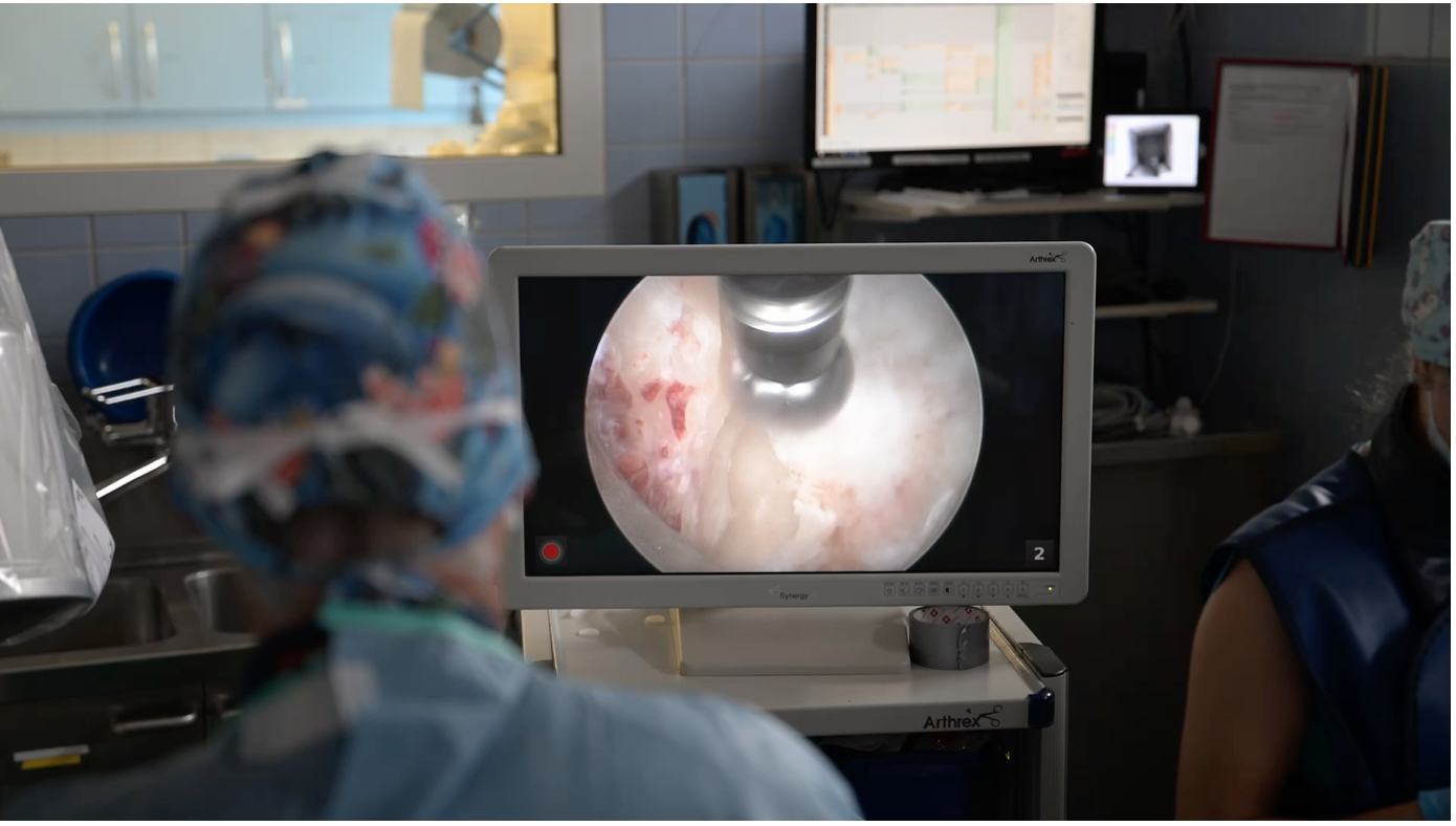 Intrasurgical view of bone being removed by a drill. Photo © private collection.
Impinged cervical nerves can be diagnosed by CT (myelography) under general anesthesia and different neck positions like a flexed or extended neck carriage can be imitated to get a better understanding of the effect of different head and neck positions on symptoms.
“You can only diagnose a lameness originating from the region of the neck by excluding other causes in the fore limb,” Swagemakers explains. “Similar to blocks in the distal limb you can block the facet joints by injecting local anesthetic, however this is not possible for the exiting nerves. If the horse presents with symptoms that are indicative of a neck issue, X-rays are the first diagnostic step giving a first impression of the cervical spine. Should there be suspicion of nerve compression due to narrowing of the foraminal space, a CT scan is necessary to make a final diagnosis. By now, we've done over 400 surgeries, so we feel more confident in our case selection than we did in the beginning and owner feedback has further improved our understanding.”
“After unsuccessful prior treatment or recurring severe symptoms, surgery is an option,” Swagemakers explains. “A CT scan is always needed to make the diagnosis and plan the surgical approach. After surgery, horses return to work under saddle after six weeks barring complications. Movement is introduced gradually with two days of box rest directly after surgery, followed by five days of hand-walking before the horses can be lunged on a halter, progressively adding different proprioceptive aids.”
Opening the nerve pathway
-
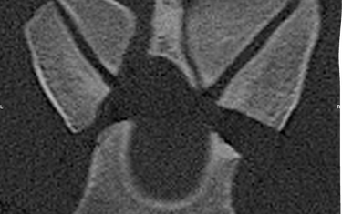
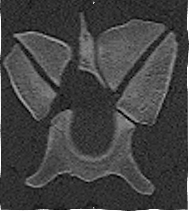
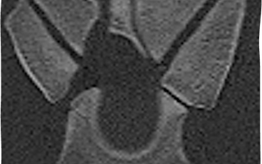 Crossection C67 before surgery with the narrowed intervertebral foramen shown
-
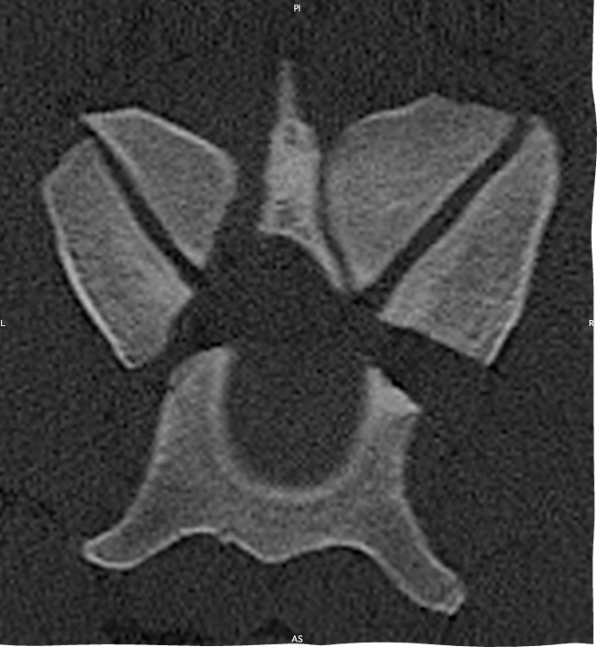
 The same image after surgery
The goal of the operation is to open up the pathway for the nerve by drilling away the bony changes causing the narrowing of the intervertebral foramen. “We access the edge of the enlarged facet ultrasound-guided with a needle first. We guide the sleeve over the needle, through which we then enter the endoscope. After removing the soft tissue, we can drill away the excess bone compressing the nerve,” Swagemakers explains. “With the spinal cord only millimetres away, supplied by major blood vessels, there is no room for error.”
Swagemakers got the idea from humane medicine. “My father-in-law got operated on his lumbar spine because he had sensory deficits, i.e. tingling, in his toes,” he explains. “When my mother-in-law told me that they were going to operate his spine by a minimally invasive technique, similar to an arthroscopy, I was very sceptical. In a joint, you can go in and distend the joint with gas or fluid so you have space to work and visualize, but that’s not possible in the spine. As I was curious how they were going to do the surgery, I went to the hospital and was allowed to watch the whole procedure.”
Inspired, Swagemakers returned to his clinic in Germany. “Because in the human field these surgeries are mostly performed in the lumbar spine, we started evaluating a number of lumbar CT images, but as it turns out, we see more pathology in the cervical spine in horses,” he recalls. “We collaborated with a human neurosurgeon and performed the first surgery in 2020. Since then, the technique has advanced quite a bit and remains a safe and effective method to address cervical nerve issues”
22.1.2026 No reproduction of any of the content in this article will be accepted without a written permission, all rights reserved © World of Showjumping.com. If copyright violations occur, a penalty fee will apply.